
Medical and Health Science HeartAnatomy!!
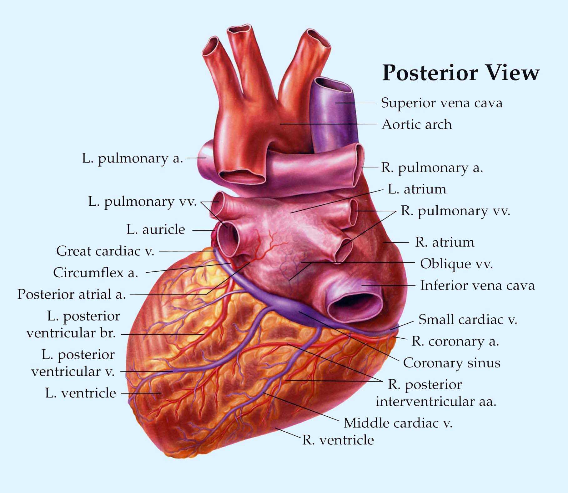
Definition. enlarged vessel on the posterior aspect of the heart that empties blood into the right atrium. Location. Term. Posterior vein of left ventricle. Definition. Parallels the posterior left ventricular branch. Location.

Posterior view of adult human heart showing distribution of cardiac
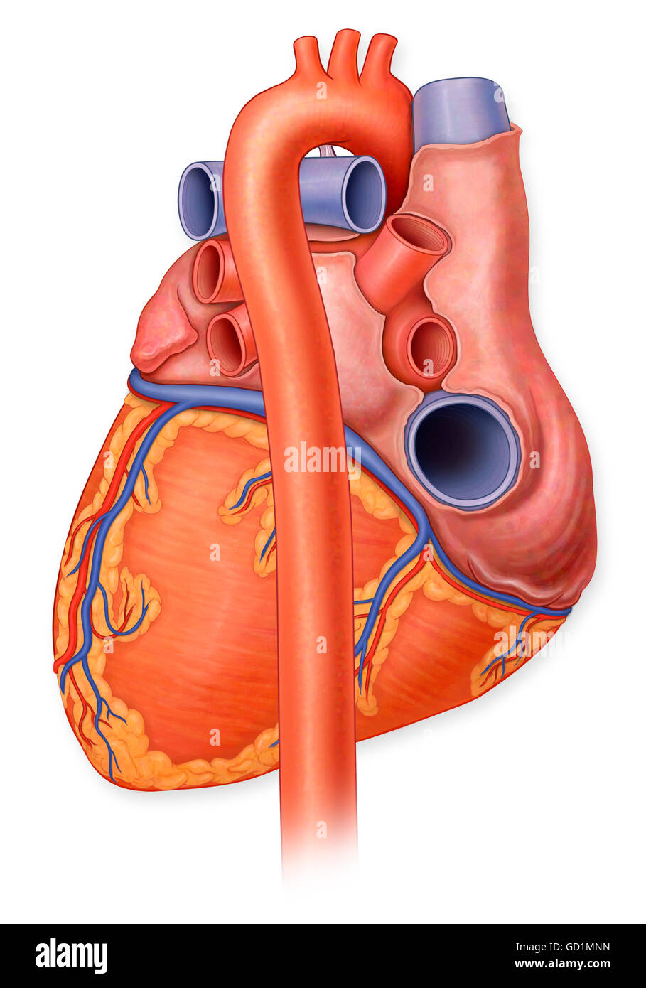
Heart, posterior view. View Media Gallery The overall shape and position of the heart may vary according to the relative size and orientation of each of its parts. For example, a large.
Human Heart Posterior View Carlson Stock Art
Location of the Heart (Posterior View) 3. 8. Hide the lungs by deselecting the respiratory system icon on the left-hand side of the screen.. Coronary Circulation (Posterior View) 11. a. Select the right coronary artery. This artery and its branches supply blood to the _____ and the _____. It extends from the _____ and runs to the right of.

Heart Anatomy Anatomy and Physiology II
Figure 1. The heart is located within the thoracic cavity, medially between the lungs in the mediastinum. It is about the size of a fist, is broad at the top, and tapers toward the base. Everyday Connection: CPR
Posterior view of a normal heart and it's arteries Stock Photo Alamy
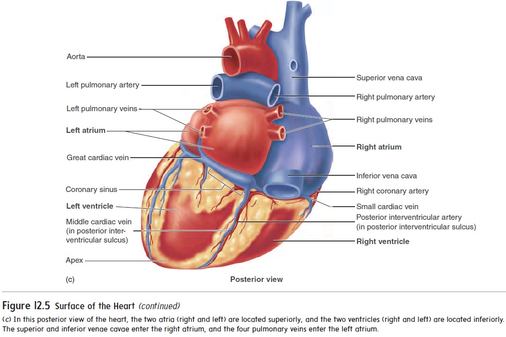
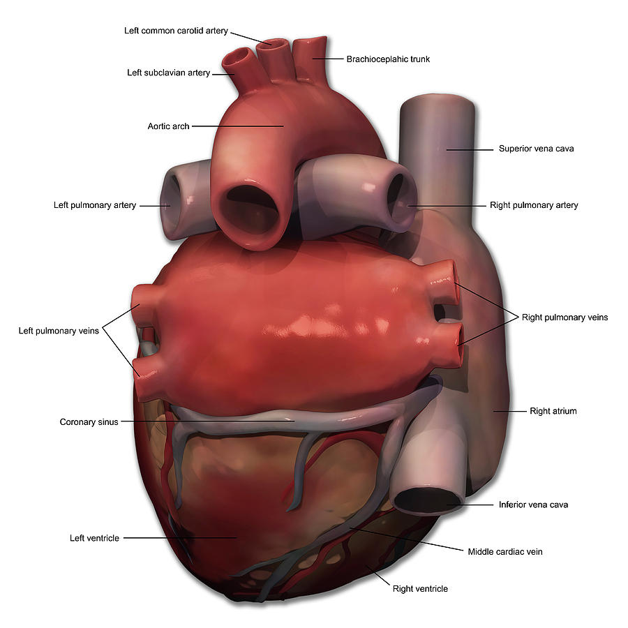
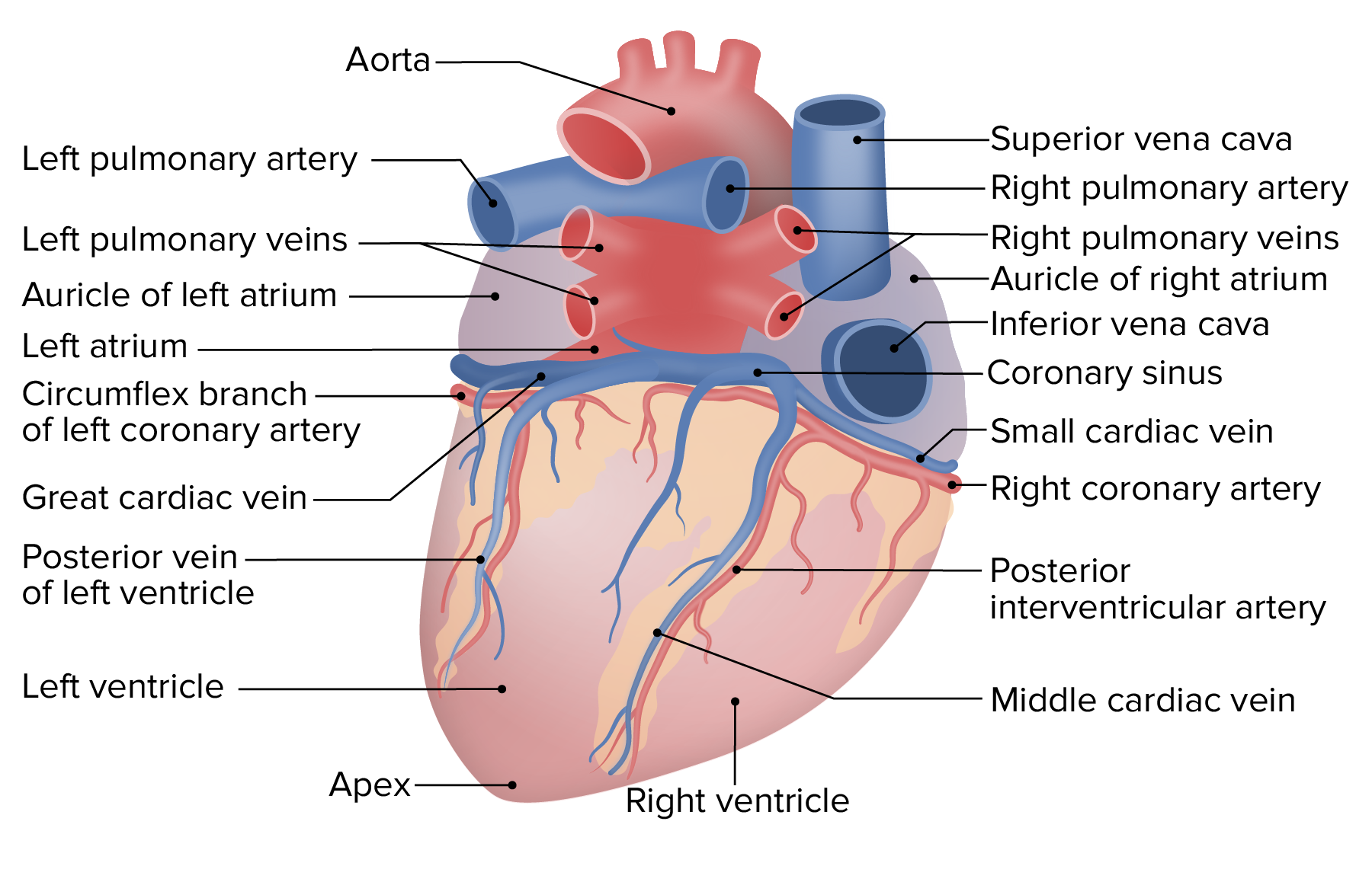
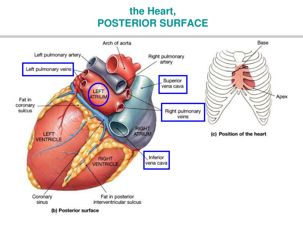
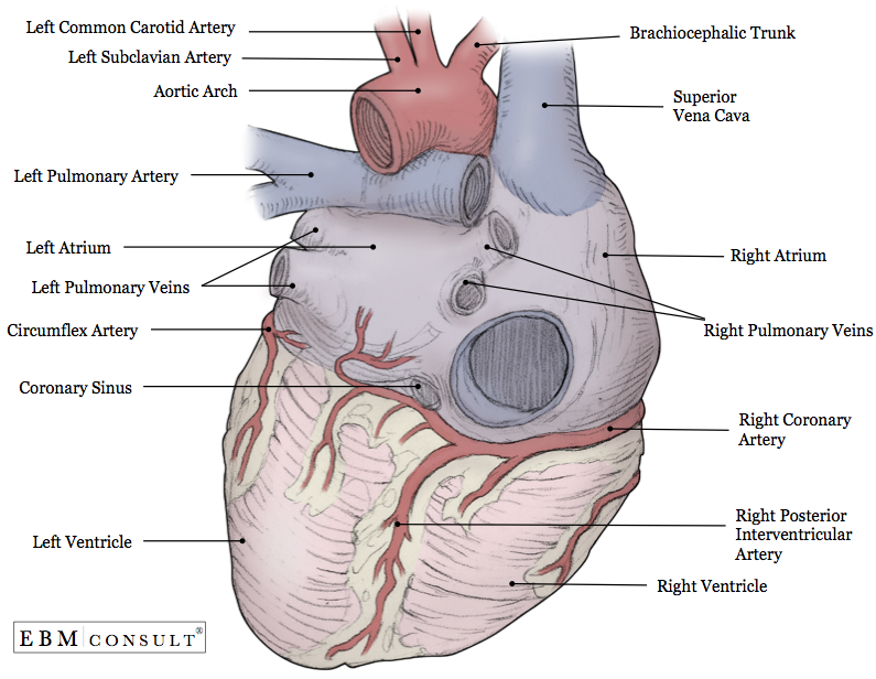
Heart anatomy The heart has five surfaces: base (posterior), diaphragmatic (inferior), sternocostal (anterior), and left and right pulmonary surfaces. It also has several margins: right, left, superior, and inferior: The right margin is the small section of the right atrium that extends between the superior and inferior vena cava .
Anatomy of the Heart
Anatomy of the Heart. Figure 1. The endocast is viewed from 5 different perspectives to demonstrate the spatial relationship between right (coloured blue) and left (coloured red) heart chambers and between atria and ventricles. The blue and white arrows represent the right and left ventricular outflow tracts respectively.
Heart anatomy Page 7
Learn about the significance of posterior heart view for your cardiovascular health and how Nao Medical's cardiology specialists can help you maintain a healthy heart.
Posterior View Of Human Heart Anatomy Photograph by Alayna Guza Pixels
[Figure, Posterior View of Heart and.] - StatPearls - NCBI Bookshelf StatPearls [Internet]. Show details Posterior View of Heart and Lungs, Entrance of Vena Azygos, Branch of Pulmonary Artery, Left Ventricle, Left Atrium, Great Coronary Vein. Contribute by Gray's Anatomy Plates From: Anatomy, Thorax, Lungs

4 posterior view of the human heart Download Scientific Diagram
(a) Right lateral view and (b) left lateral view. DA, descending aorta. Fig. 12.6 Heart margins in lateral projections. Cross-sections at three levels. The true posterior surface of the heart is commonly referred as the base of the heart which is formed largely by the left atrium.
Heart Anatomy Concise Medical Knowledge
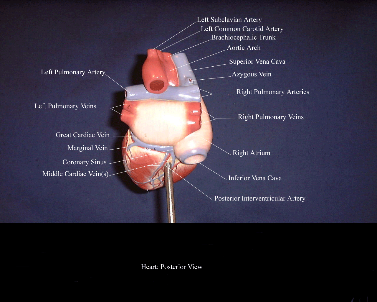
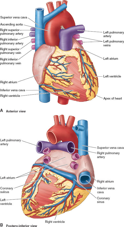
What is the Posterior View of the Heart? The posterior view of the heart is the view of the heart from the back. It shows the heart's right and left atria, right and left ventricles, and the major blood vessels that enter and exit the heart. Why is Understanding the Posterior View of the Heart Important?
PPT HEART ANATOMY PowerPoint Presentation, free download ID6415138
Figure 1. Anterior view of the heart A with and B without coronary fat and vessels. Figure 2. Posterior view of the heart A with and B without coronary fat and vessels. Figure 3. Lateral view of the right atrium of the heart, with A the outer wall of the right atrium dissected and peeled back.

Faculty and Staff Personal Web Space North Seattle Community College
Heart (Posterior View) Main Chambers of the Heart. Right Atria: Receives venous (or deoxygenated) blood from the superior and inferior vena cava and the coronary sinus and transfers. Functions to transfer blood thru the tricuspid valve during diastole (ventricular relaxation) into the right ventricle.
Anatomy Heart (External)
Location of the Heart. The human heart is located within the thoracic cavity, medially between the lungs in the space known as the mediastinum. Figure 19.2 shows the position of the heart within the thoracic cavity. Within the mediastinum, the heart is separated from the other mediastinal structures by a tough membrane known as the pericardium.
posteriorheart1
The heart has three layers. They are the: Epicardium: This thin membrane is the outer-most layer of the heart. Myocardium: This thick layer is the muscle that contracts to pump and propel blood.

Photorealistic image of the posterior view of the human heart. Four
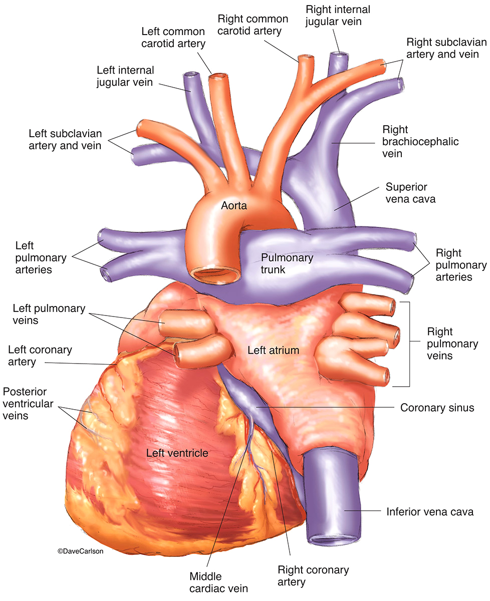
This illustration demonstrates a posterior view of the thoracic cavity, highlighting the position of the heart in relationship to the ribs and diaphragm. The left atrium, left ventricle, and coronary sinus in the coronary sulcus between these chambers can be easily seen from the posterior aspect.
Cardiovascular Anatomy and Physiology Anesthesia Key
Discover the Posterior View of the Heart: A Comprehensive Guide Learn about the posterior view of the heart and how it functions. Discover how Nao Medical can help you maintain a healthy heart. Skip to content Nao Medical After Hours service is currently available! Make an Appointment Our Services Our Services Primary care Urgent care